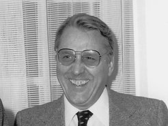
Obituary for Elmar Zeitler
The former director of the Fritz Haber Institute, Prof. Dr Elmar Zeitler, enabled many advances in electron microscopy. In December 2020, he sadly passed away. Prof. Dr. Robert Schlögl remembers him as an uncompromising yet modest scientist, and looks back on his scientific achievements.

Prof. Elmar Zeitler passed away in Berlin on 19 December 2020. He was a scientific member and director at the Fritz Haber Institute from 1977 until his retirement in 1995. Elmar was a full-blooded physicist and always interested in quantitative and analytical solutions to problems. The traceability of a problem to fundamental facts of physics and optics was always his essential concern. At the same time, Elmar was imbued with the desire to help others solve their practical problems with his fundamental contributions. He was an enabler of numerous advances in electron microscopy. He succeeded particularly well in this through his personal way of always putting the matter and never his own person in the foreground.
After finishing his PhD in 1953 in Würzburg on the subject of "Investigations on the hard secondary radiation of cosmic rays", he worked until 1958 at Bayer in Leverkusen on the knowledge-based perfection of scientific photography. A research stay at the Karolinska Institutet in Stockholm in 1958 awakened his interest in the electron microscopy of biological objects, a subject that would accompany him for the rest of his life. After his return to the University of Würzburg, where he achieved habilitation in 1960, he gave the lecture "Physics for Physicians". During this time, he developed a method to determine the local mass of a (biological) object by means of quantitative contrast analysis. This method could easily have been achieved with modern microscopes, but was an experimental feat in its time, for which Elmar also created the precise analytical basis.
This was followed by his "American phase" at Walter Reed Hospital in Washington and as a professor at the Department of Physics and Biophysics at Chicago University, where he worked until 1977. During this time, the professor of physics was concerned with radiation damage to medical specimens and the need for digital reconstruction. In this capacity, Elmar Zeitler imparted his profound knowledge of optics and the mathematical description of the interaction of electrons and matter to numerous students who later adopted his methodology in their own research. Scientifically, he was involved in the development of scanning transmission microscopy (STEM) and the point electron source (field emission method) required for it. He developed a self-built field emission source on the basis of a commercial electron microscope. At the same time, he generated software for image reconstruction on a central computer based on his work on the analytical description of the 3-dimensional reconstruction of 2-dimensional images. A publication on haemoglobin and sickle cell anaemia ("Electron microscopy of fibres and discs of HemoglobinS having sixfold symmetry", Proc. Natl. Acad. Sci. USA, vol 74, (1977) p. 5538) showed the potential of this method. This work is characteristic of the work of Elmar Zeitler, who was simultaneously involved in all aspects of microscopy, from sample preparation and imaging methodology to the development of instrumentation and digital data analysis.
In 1977, Elmar Zeitler was appointed to succeed Ernst Ruska at the Fritz Haber Institute. With the additional experimental possibilities of the department of Ernst Ruska, who remained at the Institute for a long time as an emeritus professor, Elmar was able to implement his ideas of a fundamental solution to the beam damage problem and develop the spectroscopy of energy loss in the microscope with the possibilities of real space imaging as micro-spectroscopy. The fundamental concern of his work in Berlin was to master and use the interaction of high-energy electrons with matter. Their negative side are the phenomena of beam damage, which makes imaging of biology-sensitive objects in particular very difficult. The positive side is the information about the local electronic structure of the imaged atoms carried by the outgoing electron beam. The answers to this challenge were cryoelectron microscopy and electron energy loss spectroscopy (EELS). The following quote shows how aware Elmar was of the fundamental importance of his work and how critical he was of his science:
In most cases, the development of a field does not follow logical lines. There are bandwagons which lead wayward; sometimes stumbling blocks are bypassed because a new direction may point to potential success. Problems are glossed over, put in abeyance or simply forgotten, repressed or whatever other psychological terms might be fitting. But as in fashion or psychology, finally they come out of the closet. One such challenge is radiation damage – (E.Z. Ultramicroscopy vol.10, 1982, p. 1)
Elmar Zeitler understood cryomicroscopy as a system of measures to organise the observation of an object so that it became visible in its native state. He utilised superconducting objective lenses to use stable magnetic fields (this is achieved today by modern electronics), he developed methods of preparing specimens in amorphous ice and he developed specimen holders for low-temperature experiments (both still in use today). For the perfect 3-D reconstruction of the projection images from the microscope, fundamental considerations of image reconstruction and digital image processing were necessary, which Elmar was able to develop in a powerful form and was thus far ahead of his time. In his own way, Elmar facilitated the dissemination of his concepts and ideas through collaborations. Industrial users commercialised his technologies. Scientists conducted corresponding pioneering experiments with the hardware and software available in Berlin. This led to the key publication for today's highly topical field of biological cryomicroscopy "Model for the Structure of Bacteriorhodopsin based on High-resolution Electron Cryo Microscopy" (J. Mol. Biol. (1990, vol 213, p. 899) by the Henderson group of Cambridge. In this paper, two of Elmar Zeitler's co-workers (F. Zemlin and E. Beckmann) are co-authors as experimenters, but not Zeitler himself, although he was behind all the machinery and conception set up at that time.
The EELS method was the method of choice for Elmar Zeitler to approach the electronic structure of light atoms (as in biological samples) and to obtain the same information as is readily available for heavier atoms by means of X-ray spectroscopy. The lengthy technical development of a spectrometer with sufficient resolution was again successfully tested in cooperation, this time with Sir John Thomas from Cambridge. However, others were experimentally more successful at this time with a technically more robust solution. The problematic unfolding of spectra from their background, on the other hand, remained a topic of Elmar Zeitler's work until the end of his scientific career.
Elmar Zeitler continued to be intensively involved in the main field of work at the Fritz Haber Institute. The then as now not well-resolved definedness of the sample environment of an electron microscope was addressed experimentally many times, but the results were limited. Much more successful was the combination of a UHV surface physics for preparation and manipulation of metal surfaces followed by transfer to an electron microscope and imaging of the surface in reflection. This method was perfected and thoroughly studied conceptually by a group of people in the Zeitler department. Had it not been for the development of scanning probe methods around the same time, this technique, which could still offer numerous advantages today, would not have been forgotten. More success was achieved with the development of a UHV-capable photoelectron emission microscope (PEEM), which, based on earlier applications by others, had excellent functional characteristics due to its robust design. This instrument enabled numerous works in all departments of the Fritz Haber Institute and was commercialized according to the "Zeitler method" by cooperation with a company and thus made available to the whole community, which still uses it intensively today. And again, Zeitler's name is missing on the corresponding key publication.
His German-American experience and cooperative nature gave Elmar Zeitler many opportunities to get involved in the electron microscopy community, which he knew both from a methodological and a large subgroup biological microscopy perspective. Elmar was famous for his many stories from his rich life, which, coming from him, always brought to life the human factor behind rigorous science. The founding of a journal "ultramicroscopy" in 1975, supported by a whole number of professional communities, is an enduring achievement - especially considering that at that time such undertakings were quite unusual and required much personal persuasion.
Elmar Zeitler was an uncompromising scientist. He demanded of himself and his staff a rigorous analysis of problems and the relentless pursuit of a path once taken, regardless of the problems that arose. There were no detours and no giving up. At the same time, Elmar Zeitler was very liberal and encouraged the coexistence of working groups in his department that dealt with very different issues, as long as they worked on them rigorously in principle. He encouraged the careers of his employees who wanted to develop themselves further and at the same time built up a team of permanently employed experts in electron microscopy who formed the methodological backbone of his research. He was scientifically active himself until the end of his working life and ran a private laboratory in which he experimented with a few collaborators. Elmar Zeitler always cultivated a cooperative working style with a very wide network of personal relationships. A tradition which he initiated is the annual New Year's reception for all employees of the Fritz Haber Institute.
Even after his retirement in 1995, Elmar Zeitler advised his former department in its further development. Electron microscopy continued to play an important role, but the science changed from methodological development to the further advancement of its use in the service of catalysis research. Elmar Zeitler made exemplary use of the opportunities offered by the Max Planck Society to implement his own scientific ideas and individual working style. His cooperative and outwardly modest manner may have detracted from the appreciation of his merits, but the scientists of electron microscopy and all those who worked with him will cherish his memory.
Prof. Dr. Robert Schlögl
(Translation from the German original)


