Looking at Surfaces with Atomic-Scale Light

In order to understand complex processes on material surfaces like heterogeneous catalysis, detailed chemical information about the composition and structure of surfaces is needed. Vibrational spectroscopy can provide this information and is already used to characterize various substances in a broad range of scientific areas from physics, chemistry, biology, to medical science. In the research of surface science and heterogeneous catalysis, vibrational spectroscopy has played a central role to identify chemical species on surfaces and to learn about microscopic reaction mechanisms at the molecular level.
While there are already methods to study these things, such as Raman spectroscopy, they lack sensitivity and spatial resolution. “Tip-enhanced Raman spectroscopy is an emerging method combining the chemical sensitivity of Raman spectroscopy with the atomic-level spatial resolution of scanning probe microscopy. Recent advancements even allow us to observe single molecules. Now, a research team at the Fritz Haber Institute demonstrated a novel way to further increase the sensitivity of this spectroscopy” explains Takashi Kumagai, Research Group Leader at the Physical Chemistry Department and Center for Mesoscopic Science in Institute for Molecular Science, Japan.
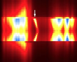
The key to all of this is precise control of light confined to nanometer scale, so-called plasmonic field, which determines the sensitivity and spatial resolution of spectroscopy. The team around Takashi Kumagai has developed techniques to use this “nanolight” which occurs at an atomically-sharp metallic tip. As the nature of such an extreme light and its interaction with materials are still largely unknown, the research team in the Department of Physical Chemistry has developed a state-of-the-art scanning probe microscope combined with laser optics and nanoplasmonics in order to explore this further. They found out something remarkable: when they applied TERS to study an oxide film on a metal surface, they discovered that the Raman scattering can be dramatically enhanced by forming atomic point contact between the tip apex and the surface. As Raman scattering contains valuable information about materials, this means that the researchers can learn more about the surface they are studying. The experiment furthermore showed that this dramatic increase strongly depends on the chemical properties of the atomic point contact. This finding will further expand the possibility of TERS to investigate atomic-scale structures and the chemistry of nanosystems.
“It has only recently been found that light can be confined like this. Our perspective is to know much more about the intriguing interaction between this extreme light and matter on the atomic level using the new scanning probe microscope” explains Takashi Kumagai. This method has great potential for ultra-sensitive and high spatial resolution spectromicroscopy, and will likely enable scientists to study as-yet-unknown photo-physics and photo-chemistry in the nanoworld.


