CECAM Flagship Workshop: From operando electron microscopy images to atomistic models: Machine Learning assisted analysis in the age of big data
- Start: Jul 2, 2025
- End: Jul 4, 2025
- Location: Zuse Institute, Takustraße 7, 14195, Berlin
- Room: Seminarraum

The advent of in situ and operando analytical techniques in material
science has allowed uncovering the importance of structural dynamics in
research fields such as heterogeneous catalysis [1], nucleation and
growth of crystals [2] or proteins [3]. This holds, in particular for
electron microscopy related operando approaches, true to the slogan:
“seeing is believing" [4]. In general, operando electron microscopy experiments cause relatively
large data stacks, for which the analysis per se is time consuming and
the statistical relevance and quantitative analysis is often missing.
Consequently, a correlation of important features with the function is
often impossible. In addition, the interaction of the highly energetic
electrons with the liquid or gaseous environment forms reactive radicals
that can interact with the matter under study and alter its atomistic
structure [5]. Thus, a true atomistic description, which can be obtained
from high resolution ex situ analysis [6], is not directly possible.
However, this knowledge would be required to understand the origin of
the function of the investigated material. Consequently, to reduce the
harmful electron-environment interaction to a negligible minimum often
operando electron microscopy experiments are conducted at lower
resolution or with a bad signal-to-noise ratio [4].On the theory side,
ML-enabled techniques have recently allowed for unprecedented modelling
of complex functional materials systems by molecular dynamics and
advanced sampling techniques [7]. Compared to biophysical simulations of
e.g. molecular machines, detailed experimental structural data that
would allow for in-depth validation of models of interfaces in catalysis
or energy-conversion systems is largely missing and urgently
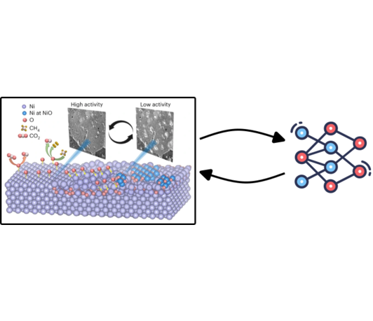
required.To overcome the outlined dilemma, machine learning aided
computervision approaches have been identified to be of outmost
importance in enhancing the interpretative depths of operando electron
microscopy experiments [8-10]. In addition, they are able to generate
automated atomistic models from huge operando electron microscopy data
stacks recorded at low resolution or signal-to-noise ratio. Atomistic
simulations can aid in detecting unlikely outliers or act as
reqularization to such approaches, building on related ideas that e.g.
allowed for NMR structure prediction of non-crystalizable proteins.The
applied machine learning approaches rely on neural network algorithm,
such as convolution neural network (CNN), fully convolution network
(FCN) or U-Net [8], which enable novel insights via automated analysis
into defects, structure, morphology and spectral features. Consequently,
linking electron microscopy to integrated machine learning and
atomistic simulation approaches enhances not only the quantitative and
statistical expression and the temporal evolution of the feature under
study, but also the generation of realistic atomistic models towards a
more accurate simulation of the function of the material under study.
For instance, CNN assisted data evaluation has generated a 3D atomistic
model of nanoparticles with the precise determination of the thickness
[11]. In addition, applying a U-Net algorithm for Au nanoparticles
segmentation during an in situ transmission electron microscopy (TEM)
experiment has disclosed the curvature dependent edging rate of these
particles [12]. To ultimately automatize experiments, recent advances in
interpretable machine learning can be of direct impact [13]. These
architectures can permit to discover governing equations, which could
serve for controlling experimental apparatus [14].
[1] J. Thomas, K. Zamaraev, Top. Catal., 1, 1-8 (1994)
[2] Z. Wang, C. Liu, Q. Chen, Journal of Crystal Growth, 601, 126955 (2023)
[3] E. Sisley, E. Illes-Toth, H. Cooper, TrAC Trends in Analytical Chemistry, 124, 115534 (2020)
[4] S. Chee, T. Lunkenbein, R. Schlögl, B. Roldán Cuenya, Chem. Rev., 123, 13374-13418 (2023)
[5] N. Schneider, M. Norton, B. Mendel, J. Grogan, F. Ross, H. Bau, J. Phys. Chem. C, 118, 22373-22382 (2014)
[6]
H. Türk, F. Schmidt, T. Götsch, F. Girgsdies, A. Hammud, D. Ivanov, I.
Vinke, L. de Haart, R. Eichel, K. Reuter, R. Schlögl, A. Knop‐Gericke,
C. Scheurer, T. Lunkenbein, Adv. Materials. Inter., 8, (2021)
[7] H. Jung, L. Sauerland, S. Stocker, K. Reuter, J. Margraf, npj. Comput. Mater., 9, 114 (2023)
[8]
S. Kalinin, D. Mukherjee, K. Roccapriore, B. Blaiszik, A. Ghosh, M.
Ziatdinov, A. Al-Najjar, C. Doty, S. Akers, N. Rao, J. Agar, S.
Spurgeon, npj. Comput. Mater., 9, 227 (2023)
[9] Z. Cheng, C. Wang, X. Wu, J. Chu, J. Semicond., 43, 081001 (2022)
[10] M. Botifoll, I. Pinto-Huguet, J. Arbiol, Nanoscale Horiz., 7, 1427-1477 (2022)
[11] M. Ragone, V. Yurkiv, B. Song, A. Ramsubramanian, R. Shahbazian-Yassar, F. Mashayek, Computational Materials Science, 180, 109722 (2020)
[12] L. Yao, Z. Ou, B. Luo, C. Xu, Q. Chen, ACS Cent. Sci., 6, 1421-1430 (2020)
[13] S. Brunton, J. Proctor, J. Kutz, Proc. Natl. Acad. Sci. U.S.A., 113, 3932-3937 (2016)
[14] M. Hoffmann, C. Fröhner, F. Noé, The Journal of Chemical Physics, 150, (2019)